Table of Contents
1. Introduction to Internal Decapitation 2. Anatomy and Physiology Involved 3. Causes and Risk Factors 4. Identifying Symptoms and Signs 5. Diagnostic Procedures 6. Emergency Response and Initial Treatment 7. Surgical Interventions 8. Post-operative Care and Rehabilitation 9. Prognosis and Recovery Outcomes 10. Case Studies and Real-life Examples 11. Preventive Measures and Recommendations 12. Advances in Medical Research 13. Psychological and Emotional Impact 14. Support Systems and Resources 15. Frequently Asked Questions 16. ConclusionIntroduction to Internal Decapitation
Internal decapitation, medically termed atlanto-occipital dislocation, is an injury where the ligaments connecting the skull to the spine are severely damaged or severed. Unlike external decapitation, there is no visible separation of the head from the body, making it a silent yet potentially fatal condition. The rarity of this injury contributes to its mystique and the critical need for awareness and rapid response.
The condition primarily affects the junction between the skull and the cervical spine, specifically the atlas (C1) and axis (C2) vertebrae. This region is crucial for supporting the head and allowing its range of motion. When the connection between the skull and spine is compromised, it can lead to significant neurological deficits or death if not promptly addressed.
Understanding internal decapitation requires a deep dive into the anatomy involved, the mechanisms leading to such injuries, and the medical protocols in place to handle them. This knowledge not only aids medical professionals in diagnosis and treatment but also empowers individuals to recognize the signs and seek timely medical help.
Anatomy and Physiology Involved
The human cervical spine is a marvel of biological engineering, designed to provide both support and flexibility. It comprises seven vertebrae, with the atlas (C1) and axis (C2) playing pivotal roles in supporting the skull and facilitating head movement. The occipital bone, located at the base of the skull, connects to the atlas, forming the atlanto-occipital joint, which is crucial for nodding motions.
This joint is stabilized by several ligaments, including the tectorial membrane and the alar ligaments. These structures are responsible for maintaining the alignment of the skull and spine, preventing excessive movement that could lead to injury. In internal decapitation, these ligaments are either stretched beyond their limits or ruptured, leading to instability in the atlanto-occipital joint.
The complexity of this region means that even minor injuries can have profound effects. Damage to the spinal cord or brainstem, which are located in close proximity, can result in neurological impairments or life-threatening conditions. Thus, a detailed understanding of the anatomy and physiology of this region is essential for both diagnosis and treatment.
Causes and Risk Factors
The causes of internal decapitation are varied and often involve high-impact trauma. Motor vehicle accidents are the leading cause, where rapid deceleration or whiplash forces can exert immense strain on the cervical spine. Other causes include falls from significant heights, sports injuries, and severe physical assaults.
Risk factors for internal decapitation include age, with children being more susceptible due to their larger head-to-body ratio and less developed musculature. Moreover, individuals with pre-existing cervical spine conditions or weakened connective tissues may be at higher risk. Understanding these risk factors is crucial for prevention and early intervention.
In recent years, there has been increased awareness of the importance of protective gear in sports and vehicles. Helmets, seat belts, and neck braces have been shown to reduce the risk of cervical spine injuries, including internal decapitation. Public health campaigns and safety regulations continue to play a significant role in mitigating these risks.
Identifying Symptoms and Signs
Recognizing the symptoms of internal decapitation is challenging due to the absence of external signs. However, certain indicators can alert medical professionals and laypersons to the possibility of this injury. Common symptoms include severe neck pain, limited range of motion, and neurological deficits such as numbness, tingling, or weakness in the limbs.
In severe cases, individuals may exhibit signs of spinal cord injury, including paralysis or respiratory distress. Loss of consciousness or altered mental status can also occur, especially if the brainstem is affected. These symptoms necessitate immediate medical evaluation and intervention.
Given the subtlety of these symptoms, it's crucial for first responders and healthcare providers to maintain a high index of suspicion when assessing individuals with potential cervical spine injuries. Early recognition and stabilization are vital to prevent further damage and improve outcomes.
Diagnostic Procedures
Diagnosing internal decapitation requires a combination of clinical assessment and advanced imaging techniques. Initial evaluation focuses on the patient's history, mechanism of injury, and presenting symptoms. Physical examination may reveal tenderness, deformity, or neurological deficits in the cervical region.
Imaging studies play a pivotal role in confirming the diagnosis. X-rays can provide an initial overview of the cervical spine alignment, but more detailed imaging is often necessary. Computed tomography (CT) scans offer a comprehensive view of the bone structures, while magnetic resonance imaging (MRI) is invaluable for assessing soft tissues, including ligaments and the spinal cord.
In cases where internal decapitation is suspected, it is essential to immobilize the patient immediately to prevent further injury. This involves the use of cervical collars or spinal boards during transport to a medical facility. Rapid and accurate diagnosis is the cornerstone of effective treatment and improved patient outcomes.
Emergency Response and Initial Treatment
The initial response to suspected internal decapitation is critical in determining the patient's prognosis. Emergency medical services (EMS) personnel play a key role in stabilizing the patient and preventing further injury during transport. Immobilization of the cervical spine is paramount, achieved through the use of cervical collars and spinal boards.
Upon arrival at a medical facility, the patient undergoes a thorough evaluation, including imaging studies to confirm the diagnosis. In cases of severe neurological impairment or instability, immediate surgical intervention may be necessary to stabilize the spine and prevent further damage.
In the emergency setting, maintaining the patient's airway, breathing, and circulation is a priority. This may involve intubation or ventilation support if there is respiratory compromise. Pain management and neuroprotection strategies are also employed to optimize outcomes.
Surgical Interventions
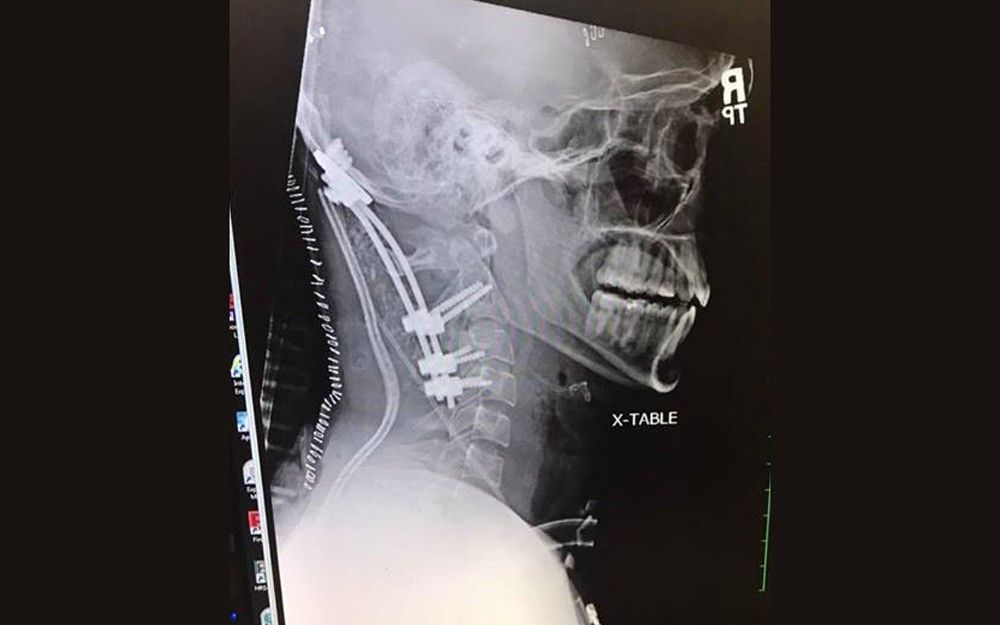
Surgery is often required to treat internal decapitation, particularly in cases where there is significant instability or neurological compromise. The primary goal of surgery is to stabilize the atlanto-occipital joint and prevent further injury to the spinal cord and brainstem.
Surgical techniques may include spinal fusion, where bone grafts and hardware such as screws and rods are used to immobilize the affected vertebrae. This procedure aims to restore stability to the cervical spine and facilitate healing. In some cases, decompression surgery may be necessary to relieve pressure on the spinal cord.
The choice of surgical intervention depends on various factors, including the severity of the injury, the patient's overall health, and the presence of any complicating factors. Surgeons work closely with a multidisciplinary team to develop a comprehensive treatment plan tailored to the patient's needs.
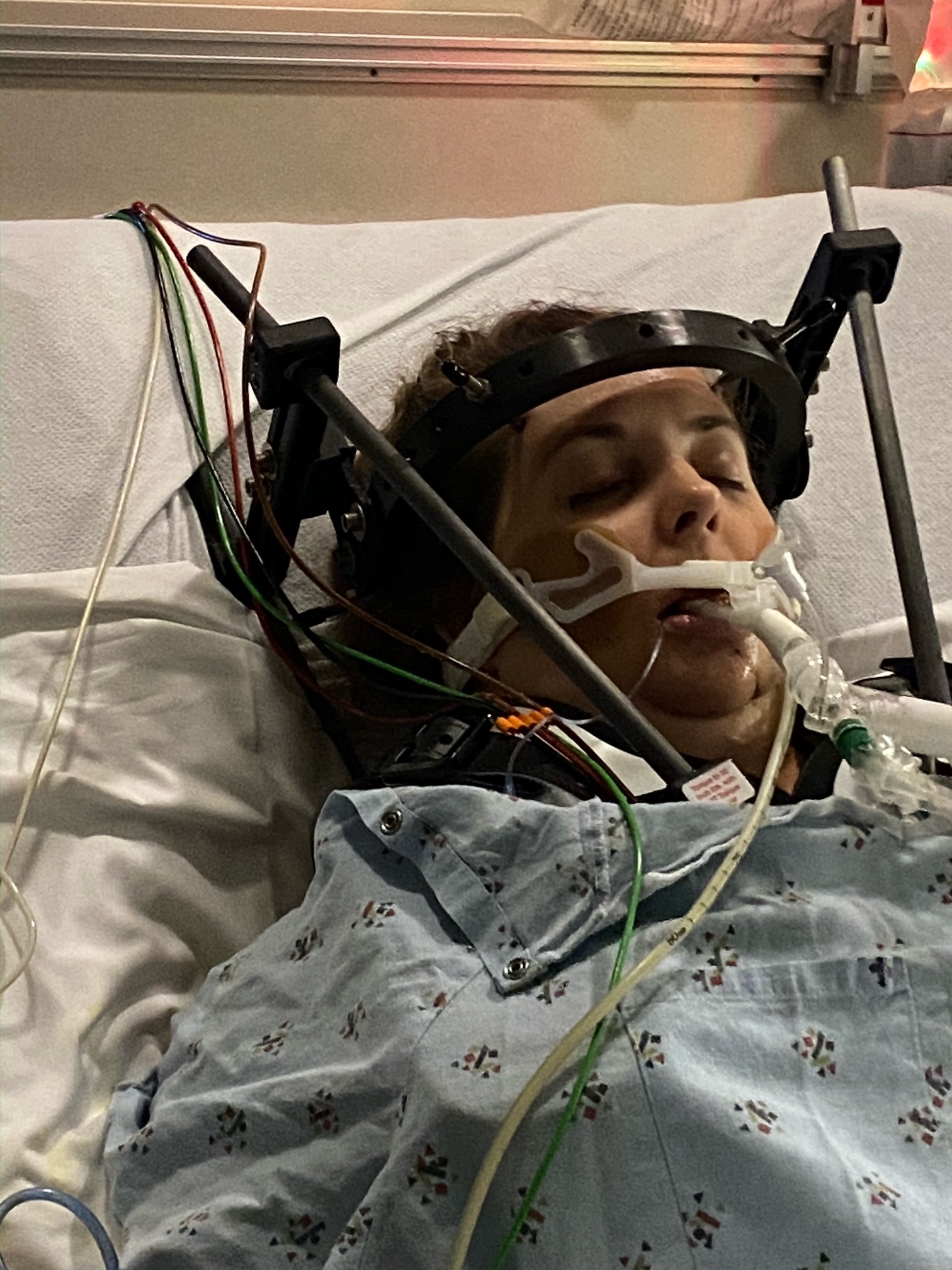
Post-operative Care and Rehabilitation
Recovery from internal decapitation is a complex and multifaceted process that requires intensive post-operative care and rehabilitation. Following surgery, patients are typically monitored in an intensive care unit (ICU) to ensure stability and address any immediate complications.
Rehabilitation plays a crucial role in the recovery process, focusing on restoring function, strength, and mobility. Physical therapy, occupational therapy, and speech therapy may be necessary to address the diverse needs of the patient. The rehabilitation plan is tailored to the individual's specific deficits and goals.
Emotional and psychological support is also vital during the recovery process. Patients may experience anxiety, depression, or post-traumatic stress disorder (PTSD) as a result of their injury and hospitalization. Access to mental health resources and support groups can significantly enhance recovery and quality of life.
Prognosis and Recovery Outcomes
The prognosis for individuals with internal decapitation varies widely and depends on several factors, including the severity of the injury, the timeliness of intervention, and the presence of any associated complications. While some patients may achieve significant recovery, others may experience long-term neurological deficits or require ongoing medical support.
Advancements in medical technology and surgical techniques have improved survival rates and outcomes for patients with internal decapitation. Early intervention and comprehensive rehabilitation are key to optimizing recovery and minimizing long-term disabilities.
It's important for patients and their families to have realistic expectations and a clear understanding of the recovery process. Regular follow-up appointments and ongoing communication with healthcare providers are essential to monitor progress and address any emerging issues.
Case Studies and Real-life Examples
Examining real-life cases of internal decapitation provides valuable insights into the challenges and triumphs associated with this condition. Each case is unique, highlighting the importance of personalized treatment plans and the resilience of the human body.
One notable case involves a young adult who survived a high-speed car accident resulting in internal decapitation. Through prompt medical intervention and a rigorous rehabilitation program, the individual achieved significant recovery, regaining mobility and independence.
These case studies underscore the importance of rapid response, multidisciplinary care, and patient determination in overcoming the challenges posed by internal decapitation. They also serve as a source of inspiration and hope for others facing similar circumstances.
Preventive Measures and Recommendations
Prevention of internal decapitation involves a combination of public awareness, safety measures, and personal precautions. Promoting the use of seat belts and airbags in vehicles, as well as helmets in sports, can significantly reduce the risk of cervical spine injuries.
Education on the importance of safe driving practices, fall prevention, and the use of protective gear is essential for individuals of all ages. Public health campaigns and community programs can play a pivotal role in disseminating this information and fostering a culture of safety.
For individuals with pre-existing cervical spine conditions, regular medical check-ups and adherence to prescribed treatments can help mitigate the risk of injury. Awareness of personal risk factors and lifestyle modifications can also contribute to prevention efforts.
Advances in Medical Research
Ongoing research in the field of cervical spine injuries continues to enhance our understanding of internal decapitation and improve treatment outcomes. Innovations in imaging techniques, surgical methods, and rehabilitation strategies are paving the way for more effective management of this condition.
Research studies focusing on the biomechanics of cervical spine injuries are providing valuable insights into the mechanisms of internal decapitation. This knowledge is instrumental in developing preventive measures and refining surgical techniques.
Collaboration between researchers, clinicians, and medical institutions is driving progress in this field, with the ultimate goal of improving patient outcomes and quality of life. Continued investment in research and innovation is essential for advancing our understanding and treatment of internal decapitation.
Psychological and Emotional Impact
The impact of internal decapitation extends beyond physical injuries, affecting patients' psychological and emotional well-being. The trauma of the injury, coupled with the challenges of recovery, can lead to a range of emotional responses, including anxiety, depression, and PTSD.
Addressing the psychological and emotional needs of patients is a critical component of comprehensive care. Access to mental health services, counseling, and support groups can provide valuable support and coping strategies for patients and their families.
Healthcare providers play a vital role in recognizing and addressing the emotional impact of internal decapitation. A holistic approach to care, integrating physical, emotional, and psychological support, can enhance recovery and improve overall well-being.
Support Systems and Resources
Support systems and resources are essential for individuals recovering from internal decapitation. These resources provide guidance, information, and emotional support to patients and their families throughout the recovery journey.
Support groups and online communities offer a platform for individuals to connect with others who have experienced similar challenges. Sharing experiences and insights can foster a sense of community and empowerment.
Healthcare providers, rehabilitation centers, and non-profit organizations also offer a range of resources, including educational materials, counseling services, and financial assistance programs. Access to these resources can significantly enhance the recovery process and improve quality of life.
Frequently Asked Questions
What is internal decapitation?
Internal decapitation, or atlanto-occipital dislocation, is a rare injury where the ligaments connecting the skull to the spine are damaged, causing instability without visible separation.
How is internal decapitation diagnosed?
Diagnosis involves clinical assessment and imaging studies, such as CT and MRI scans, to evaluate the alignment and condition of the cervical spine and surrounding tissues.
Can someone survive internal decapitation?
Yes, with timely medical intervention and appropriate treatment, individuals can survive and recover from internal decapitation, although outcomes vary based on the severity of the injury.
What are the risk factors for internal decapitation?
Risk factors include high-impact trauma, such as car accidents or falls, as well as pre-existing cervical spine conditions and weakened connective tissues.
What is the treatment for internal decapitation?
Treatment often involves surgical stabilization of the cervical spine, followed by rehabilitation to restore function and mobility. A multidisciplinary approach is essential for optimal recovery.
How can internal decapitation be prevented?
Prevention involves the use of safety measures such as seat belts, helmets, and safe driving practices, along with public awareness and education on risk factors and protective strategies.
Conclusion
Internal decapitation, while rare, is a serious medical condition that requires prompt recognition and intervention. Advances in medical technology and treatment have improved outcomes for many individuals affected by this injury. By understanding the complexities of internal decapitation, from its causes and symptoms to diagnosis and treatment, we can better prepare to address this condition and support those on their path to recovery. Through continued research, education, and collaboration, we can further enhance our ability to prevent and manage this challenging injury, offering hope and healing to those in need.